Jeremy Norman’s
HistoryofInformation.com Exploring the History of Information and Media through Timelines
Jeremy Norman’s

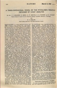
A: Cambridge, England, United Kingdom

John Kendrew and Max Perutz looking at part of the model of the structure of a protein. MRC Laboratory of Molecular Biology, Cambridge University.
In 1958 and 1960 molecular biologist John Kendrew published "A Three-Dimensional Model of the Myoglobin Molecule Obtained by X-ray Analysis" (with G. Bodo, H. M. Dintzis, R. G. Parrish, H. Wyckoff,) Nature 181 (1958) 662-666, and "Structure of Myoglobin: A Three-Dimensional Fourier synthesis at 2 Å Resolution" (with R. E. Dickerson, B. E. Strandberg, R. G. Hart, D. R. Davies, D. C. Phillips, V. C. Shore). Nature 185 (1960) 422-27. These papers reported the first solution of the three-dimensional molecular structure of a protein, for which Kendrew received the 1962 Nobel Prize in chemistry, together with his friend and colleague Max Perutz, who solved the structure of the related and more complex protein, hemoglobin, two years after Kendrew’s achievement.
Kendrew began his investigation into the structure of myoglobin in 1949, choosing this particular protein because it was “of low molecular weight, easily prepared in quantity, readily crystallized, and not already being studied by X-ray methods elsewhere” (Kendrew, “Myoglobin and the structure of proteins. Nobel Prize Lecture [1962],” pp. 676-677). Protein molecules, which contain, at minimum, thousands of atoms, have enormously convoluted and irregular formations that are extremely difficult to elucidate. In the 1930s J. D. Bernal, Dorothy Hodgkin and Max Perutz performed the earliest crystallographic studies of proteins at Cambridge’s Cavendish Laboratory; however, the intricacies of three-dimensional structure of proteins were too complex for analysis by conventional X-ray crystallography, and the process of calculating the structure factors by slide-rules and electric calculators was far too slow. It was not until the late 1940s, when Kendrew joined the Cavendish Laboratory as a graduate student, that new and more sophisticated tools emerged that could be used to attack the problem. The first of these tools was the technique of isomorphous replacement, developed by Perutz during his own researches on hemoglobin, in which certain atoms in a protein molecule are replaced with heavy atoms. When these modified molecules are subjected to X-ray analysis the heavy atoms provide a frame of reference for comparing diffraction patterns. The second tool was the electronic computer, which Kendrew introduced to computational biology in 1951. The first electronic computer, the ENIAC, which became operational in Philadelphia in 1945, was 10,000 times the speed of a human performing a calculation. In 1951 Cambridge University was one of only three or four places in the world with a high-speed stored-program electronic computer, and Kendrew took full advantage of the speed of Cambridge’s EDSAC computer, and its more powerful successors, to execute the complex mathematical calculations required to solve the structure of myoglobin. Kendrew was the first to apply an electronic computer to the solution of a complex problem in biology.
Nevertheless, even with the EDSAC computer performing the calculations, the research progressed remarkably slowly. Only by the summer of 1957 did Kendrew and his team succeed in creating a three-dimensional map of myoglobin at a resolution the so-called “low resolution”of 6 angstroms; thus myoglobin became “the first protein to be solved” (Judson, p. 538).
“A cursory inspection of the map showed it to consist of a large number of rod-like segments, joined at the ends, and irregularly wandering through the structure; a single dense flattened disk in each molecule; and sundry connected regions of uniform density. These could be identified respectively with polypeptide chains, with the iron atom and its associated porphyrin ring, and with the liquid filling the interstices between neighboring molecules. From the map it was possible to ‘dissect out’ a single protein molecule . . . The most striking features of the molecule were its irregularity and its total lack of symmetry” (Kendrew, “Myoglobin,” p. 681).
The 6-angstrom resolution was too low to show the molecule’s finer features, but by 1960 Kendrew and his team were able to obtain a map of the molecule at 2-angstrom resolution. “To achieve a resolution of 2 Å it was necessary to determine the phases of nearly 10,000 reflections, and them to compute a Fourier synthesis with the same number of terms . . . the Fourier synthesis itself (excluding preparatory computations of considerable bulk and complexity) required about 12 hours of continuous computation on a very fast machine (EDSAC II)” (Kendrew, “Myoglobin,” p. 682).